Bone Anatomy Of Ribs / Lecture 10 Axial Skeleton Ii And Appendicular Skeleton Anatomy Bones Anatomy Human Body Anatomy : However, they truly function as an organ.
Bone Anatomy Of Ribs / Lecture 10 Axial Skeleton Ii And Appendicular Skeleton Anatomy Bones Anatomy Human Body Anatomy : However, they truly function as an organ.. Ribs are highly vascular and trabecular with a thin outer layer of compact bone. It is made up of 12 pairs of ribs. Anatomy the rib cage is a bony structure found in the chest (thoracic cavity). However, they truly function as an organ. The ribs are a set of twelve paired bones which form the protective 'cage' of the thorax.
Structure of the ribcage and ribs. The angle is the area of greatest rib curvature and forms the largest portion of the thoracic cage. Rib bone anatomy quiz for students taking anatomy and physiology! In adults, red marrow is limited to the spongy bone in the skull, ribs, sternum, clavicles, vertebrae and pelvis. Like other organs, bones are valuable and have many functions.
The Thoracic Cage Physiology Americorps Health from www.americorpshealth.biz Introduction to the structure of the ribcage and ribs: There are twelve pairs of ribs, all of which articulate with the vertebral column. The rib tubercle articulates with the transverse process of a thoracic vertebra. The remaining ribs are typical. Anatomy, histology and elemental profile of long bones and ribs of the asian elephant (elephas maximus) anat sci int. You will also find the xiphoid process, 10th rib, the apex of the heart, the coronary vein of the heart. In this image, you will find clavicle, true ribs, sternal angle, costal cartilage, false ribs, floating ribs, seventh cervical vertebra, first thoracic vertebra, jugular notch, manubrium, body, xiphoid process, sternum in it. They make up the lateral part of our body, its anterior and posterior wall and they entirely build the lateral parts of the chest wall.
It is made up of 12 pairs of ribs.
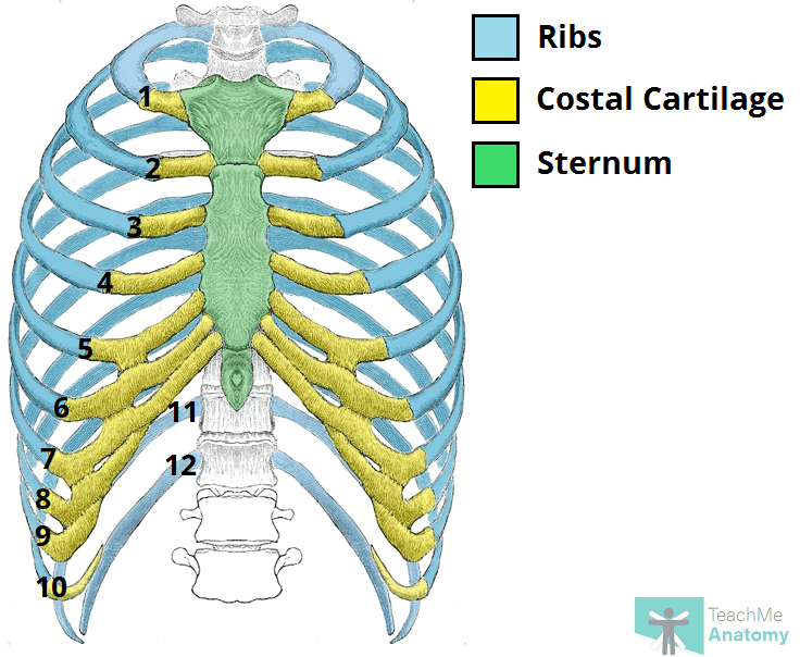
They articulate with the vertebral column posteriorly, and terminate anteriorly as cartilage (known as costal cartilage). In this image, you will find clavicle, true ribs, sternal angle, costal cartilage, false ribs, floating ribs, seventh cervical vertebra, first thoracic vertebra, jugular notch, manubrium, body, xiphoid process, sternum in it. The anatomy of the human ribs (costae) are one of the integral parts of the chest wall; Like other organs, bones are valuable and have many functions. The 1 st, 11 th and 12 th ribs are considered atypical ribs due to their anatomical features. Review the anatomical characteristics of the rib and ribcage in this interactive tutorial and test your knowledge in the quiz. They are extremely light, but highly resilient; The head of a rib is attached posteriorly to the costal facets of the thoracic vertebrae. Its function is to elevate the ribs. The human rib cage is made up of 12 paired rib bones; Ribs are highly vascular and trabecular with a thin outer layer of compact bone. The sternum is located in the center of the anterior thoracic wall and is also known as the breastbone. You may also find the sternal end, body, head, neck, tubercle, articular facets.
Chest bone, ribs, lung, heart, xiphoid process, sternum anatomy. The first 10 ribs on either side of the body have bars of hyaline cartilage, called costal cartilage.this cartilage stretches from each rib's bony end to a structure called the sternum, which is. The cartilage strips are called costal cartilage (costal is the anatomical adjective that refers to the rib) and connect on their other end to the sternum. There are 206 bones in the human skeleton, not including teeth and sesamoid bones (small bones found within cartilage): This includes the head, facial, hyoid, auditory, trunk, ribs, and sternum.
The Ribs Rib Cage Articulations Fracture Teachmeanatomy from teachmeanatomy.info The head of a rib is attached posteriorly to the costal facets of the thoracic vertebrae. Pectoral girdle and upper limb the pectoral girdle connects the upper limb (arm) bones to the axial skeleton and consists of the left and right clavicles and left and right scapulae. Lateral view of a pair of ribs articulating with the thoracic vertebrae. You may also find the sternal end, body, head, neck, tubercle, articular facets. The rib below that is rib 2, and it connects to the t2 thoracic vertebra, and so on. Introduction to the structure of the ribcage and ribs: The ribs and sternum make up what is called the 'ribcage.' the ribcage protects the lungs, blood vessels, and heart,. Anatomy the rib cage is a bony structure found in the chest (thoracic cavity).
Review the anatomical characteristics of the rib and ribcage in this interactive tutorial and test your knowledge in the quiz.
Of all 24 ribs, the first seven pairs are often labeled as 'true.' these bones are. In this image, you will find common carotid arteries, internal jugular vein, subclavian artery, subclavian vein, heart, right lung, 6th rib, diaphragm, costal cartilage in it. Instead, anatomists classify the ribs as flat bones, and they are located within the axial skeleton. They make up the lateral part of our body, its anterior and posterior wall and they entirely build the lateral parts of the chest wall. Anatomy the rib cage is a bony structure found in the chest (thoracic cavity). There are 206 bones in the human skeleton, not including teeth and sesamoid bones (small bones found within cartilage): They are extremely light, but highly resilient; You will also find the xiphoid process, 10th rib, the apex of the heart, the coronary vein of the heart. Its function is to elevate the ribs. Each pair is numbered based on their attachment to the sternum, a bony process at the front of the rib cage which serves as an anchor point. Ten of the twelve ribs connect to strips of hyaline cartilage on the anterior side of the body. Like other organs, bones are valuable and have many functions. The first 10 ribs on either side of the body have bars of hyaline cartilage, called costal cartilage.this cartilage stretches from each rib's bony end to a structure called the sternum, which is.
Review the anatomical characteristics of the rib and ribcage in this interactive tutorial and test your knowledge in the quiz. There are twelve (12) pairs of ribs and all articulate posteriorly with the thoracic vertebrae. The angle is the area of greatest rib curvature and forms the largest portion of the thoracic cage. They're cut from the upper portion of the full rib bone closer to the spine and are known in the meat industry by their more technical name, back ribs. the north american meat processors. Ribs are highly vascular and trabecular with a thin outer layer of compact bone.
Rib Cage Skeleton Thorax Heart Rib Cage Heart Experiment Cage Png Pngwing from w7.pngwing.com You will also find the xiphoid process, 10th rib, the apex of the heart, the coronary vein of the heart. The ribs are curved, flat bones which form the majority of the thoracic cage. This includes the head, facial, hyoid, auditory, trunk, ribs, and sternum. Like a house is built around a supportive frame, a strong skeleton is required to support the rest of the human body. The first 10 ribs on either side of the body have bars of hyaline cartilage, called costal cartilage.this cartilage stretches from each rib's bony end to a structure called the sternum, which is. Instead, anatomists classify the ribs as flat bones, and they are located within the axial skeleton. Of all 24 ribs, the first seven pairs are often labeled as 'true.' these bones are. Bones are often thought of as static structures which only offer structural support.
In this rib bones anatomy quiz, you can test your knowledge of the ribs.
The remaining ribs are typical. As part of the bony thorax, the ribs protect the internal thoracic organs. Without bones, it would be difficult for your body to keep its shape and to stand upright. Lateral view of a pair of ribs articulating with the thoracic vertebrae. The intermediate muscles connect to the ribs and. However, they truly function as an organ. In this image, you will find common carotid arteries, internal jugular vein, subclavian artery, subclavian vein, heart, right lung, 6th rib, diaphragm, costal cartilage in it. The 1 st, 11 th and 12 th ribs are considered atypical ribs due to their anatomical features. The angle is the area of greatest rib curvature and forms the largest portion of the thoracic cage. It acts to depress the ribs. Ribs 11 and 12 are also false ribs, but are also considered to be floating ribs because they do not have any cartilage attachment to the sternum at all. The sternum is located in the center of the anterior thoracic wall and is also known as the breastbone. If you want to learn comparative anatomy of skull, mandible, ribs and sternum among different animals then you are in right place.today i am going to discuss on comparative anatomy of skull, mandible, ribs and sternum from different animal in a short form (especially from.
This includes arms, shoulders, wrists, hands, legs, hips, ankles, and feet anatomy of ribs. Like a house is built around a supportive frame, a strong skeleton is required to support the rest of the human body.


0 Komentar